| » Behavior of osteoblasts on polymeric gratings |
| » Nanotextured self-rolled polymer micro-tubes |
Behavior of osteoblasts on polymeric gratings |
| Prof. Albert F. Yee Prof. Andy Putnam |
co-PI co-PI |
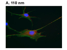 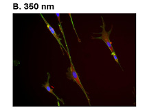 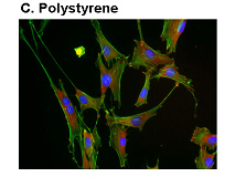 |
Studies have reported that microscale roughness and topography affect mesenchymal and osteoblast cell function. Mesenchymal cells were found to express osteoblast differentiation factors (Cfba1 and osteocalcin) significantly on grooved and rough titanium surfaces compared to tissue culture plastic. The reason behind these effects is not completely understood but it is believed that microscale roughness and topography of surfaces affects both integrin-mediated adhesion and signaling which in turn affects the expression of genes dependent on integrin signalling. Nanostructures, nanotopography or nanopatterned adhesion sites have also been shown to affect osteoblast cell shape and integrin clustering and activation. This suggests that it is possible that nanostructures are able to affect mesenchymal cell differentiation and osteoblast mineralization function through integin signaling pathways1 or through cell shape. Figure 1 shows the effect of different polymeric nanostructures on the behavior of osteoblast cells. Figure 1: MC3T3-E1 cells were cultured surfaces with PMMA nanostructures A.) 110 nm gratings, B.) 350 nm gratings and C.) unpatterend polystyrene surfaces for 24 hours. Cells were fixed and stained for F-actin (green), vinculin (red) and nuclei (blue). Representative immunofluorescent photomicrographs are shown. (20X magnification for all except (A) 40x magnification). | ||
| ↑ ↑ | ↑ ↑ |
Nanotextured self-rolled polymer micro-tubes |
| Prof. Albert F. Yee |
PI |
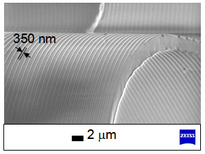 |
Nanometer features and structures have been shown to affect biological cell behavior in 2-D[1]. However, in-vivo, cells interact with nanometer textures (features and structures) in 3-D. Cell behavior in 2-D and 3-D matrices have been shown to be very different[2] so studying cell behavior in 3-D matrices is now recognized to be important. We were motivated by this trend to engineer a 3-D cell matrix with well-controlled dimensions and with nanometer textures in a reproducible way using our expertise in nanoimprint based patterning. To this end, we have successfully fabricated self-rolled polymer microtubes having nanotextures on both inner and outer surfaces. These tubes can have diameters of roughly the size of a single cell or a cluster of cells and the inner and outer surfaces of the tubes can have various nanotextures. The length of these tubes can range from several mm to several cm. In this way, the behavior of a single cell to a cluster of cells can be examined. Figure 2: SEM micrograph of micro-tube showing the nanotextures on both inner and outer surfaces. | ||
| ↑ ↑ | ↑ ↑ |